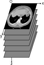
(a) Stack of 2D Slices.
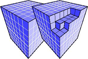
(b) Voxel Dataset.
Fig. 1: Discrete Volume Dataset.
Tomo3D-EA
Fully 3D tomographic reconstruction requires high computing power and leads to many challenges. The use of evolutionary computation in tomographic reconstruction has been largely overlooked. Tomo3D-EA is an investigation of this hypothesis in the field of in nuclear medicine. It mainly involves researchers at INRIA Saclay/Île-de-France and physicist at CEA/LIST (France). The aim is twofold: i) reduce the computing cost, and ii) produce high quality 3D dataset. Our method is based on the original fly algorithm. In practice, each fly represents a point of the object space. A fly acts as a photon or positron emitter in the case of SPECT or PET respectively. The population of fly corresponds to the object that is scanned. Initial results demonstrated favourable observations that are leading to further revisions.
Tomography
Tomography is a multi-angular analysis followed by a mathematical reconstruction. The first classical tomographic device was introduced in 1921 [3,4]. Much later, Hounsfield successfully tested the first clinical Computerized Axial Tomography (CAT, generally reduced as Computerized Tomography, or CT) in 1972 [5]. Tomographic reconstruction makes use of a large number of projections to produce a stack of trans-axial planes through the body [6]. This stack can be considered as a 3D discrete volume dataset made of voxels (see Fig. 1).
|
|
|
|
(a) Stack of 2D Slices. |
(b) Voxel Dataset. |
|
Fig. 1: Discrete Volume Dataset. |
|
Hounsfield received the Nobel Prize in Physiology or Medicine in 1979. Today, X-ray CT is still the commonest imaging modality in hospitals to routinely acquire 3D data sets from patients.
Nuclear Medicine
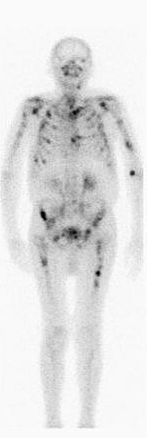
Nuclear medicine [7] appeared in the 1950's. Its principle is to diagnose or treat a disease by administering to patients a radioactive substance (also called tracer) that is absorbed by tissue in proportion to some physiological process. This is the radiolabelling process. The substance can be given by inhalation of a radioactive aerosol, ingested or, by injection of radioactive pharmaceuticals into the blood stream. In the case of diagnostic studies, the distribution of the substance in the body is then imaged: unlike in X-ray imaging or therapy, no radiation is applied from an external source. Moreover diagnostic nuclear medicine studies are generally a functional form of imaging because the purpose is to obtain information about physiological processes rather than (as with X-ray imaging) anatomical forms and structures. The photons in nuclear medicine generally have a higher energy than X-ray. Therefore dedicated detectors, called gamma camera (or Anger camera), are used instead of films. The images produced are generally referred to as scintigraphy (see Fig. 2). When a pathology occurs, the metabolism increases. There are more molecules in the pathology area. Consequently, the radioactivity also increases (see black spots in Fig. 2(b)).
|
|
|
|
(a) Normal patient. |
(b) Patient with bone cancer. |
|
Fig. 2: Bone scintigraphy. |
|
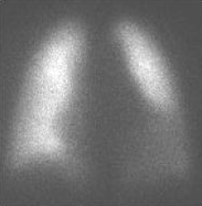
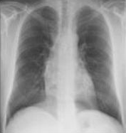
Fig. 3 illustrates that nuclear and X-ray imaging are complementary. Indeed, the first one provides information about the metabolism, in particular when it abnormally increases. The second one gives structural information about the anatomy.
|
|
|
|
(a) Lung scintigraphy. |
(b) Lung radiograph. |
|
Fig. 3: Functional versus anatomical imaging. |
|
SPECT
In the case of planar imaging, gamma cameras are used as ``films'': nevertheless it is possible to reconstruct slices through the human body using similar methods to those used in conventional X-ray computed tomography. In nuclear medicine, this method is called Single-Photon Emission Computed Tomography (SPECT) and it allows 3D reconstruction of the distribution of the tracer through the body (see Fig. 4).
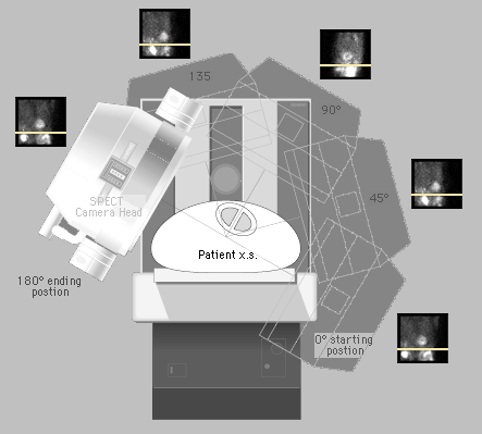
Fig.
4:
Acquisition
principle.
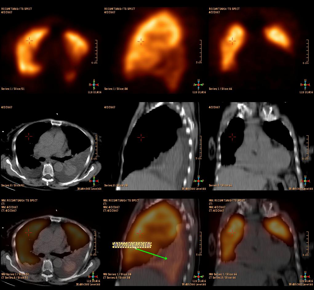
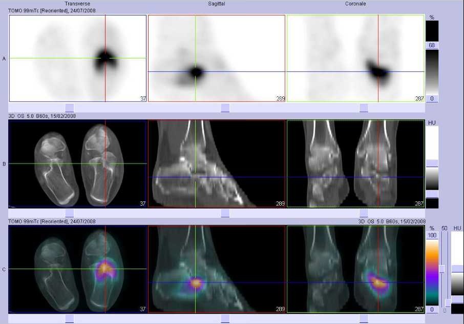
Image registration can be used to colocate SPECT and CT volumes (see Fig. 5). On the one hand, the SPECT data provides physiological information (see top rows in Fig. 5). On the other hand, the CT data provides information about the anatomical structures (middle rows in Fig. 5). The data from both imaging modalities can be superimposed (see bottom rows in Fig. 5) to combine the information.
|
|
|
|
(a) Lung with effusion of the pleura. |
(b) Foot with a fracture of the calcaneum. |
|
Fig. 5: SPECT-CT scans with i) on the top, the SPECT data, ii) in the middle, the CT data, and iii) on the bottom, the data after registration. |
|
PET
For Positron Emission Tomography (PET), a positron emitter is used as radionuclide for labeling rather than a gamma emitter. Positrons are emitted with high energy (1 MeV ). After interactions, a positron combines with an electron to form a positronium, then the electron and positron pair is converted into radiations: this is the annihilation reaction which generally produces two photons of 511 keV emitted in opposite directions. Annihilation radiations are then imaged using a system dedicated to PET containing an Anger camera specialized in high-energy. This system operates on the principle of coincidence, i.e. the difference in arrival times of the photons of each pair of detected photons and by knowing that each annihilation produces two photons emitted in exactly opposite positions: 3D images are then reconstructed in the same way as SPECT.
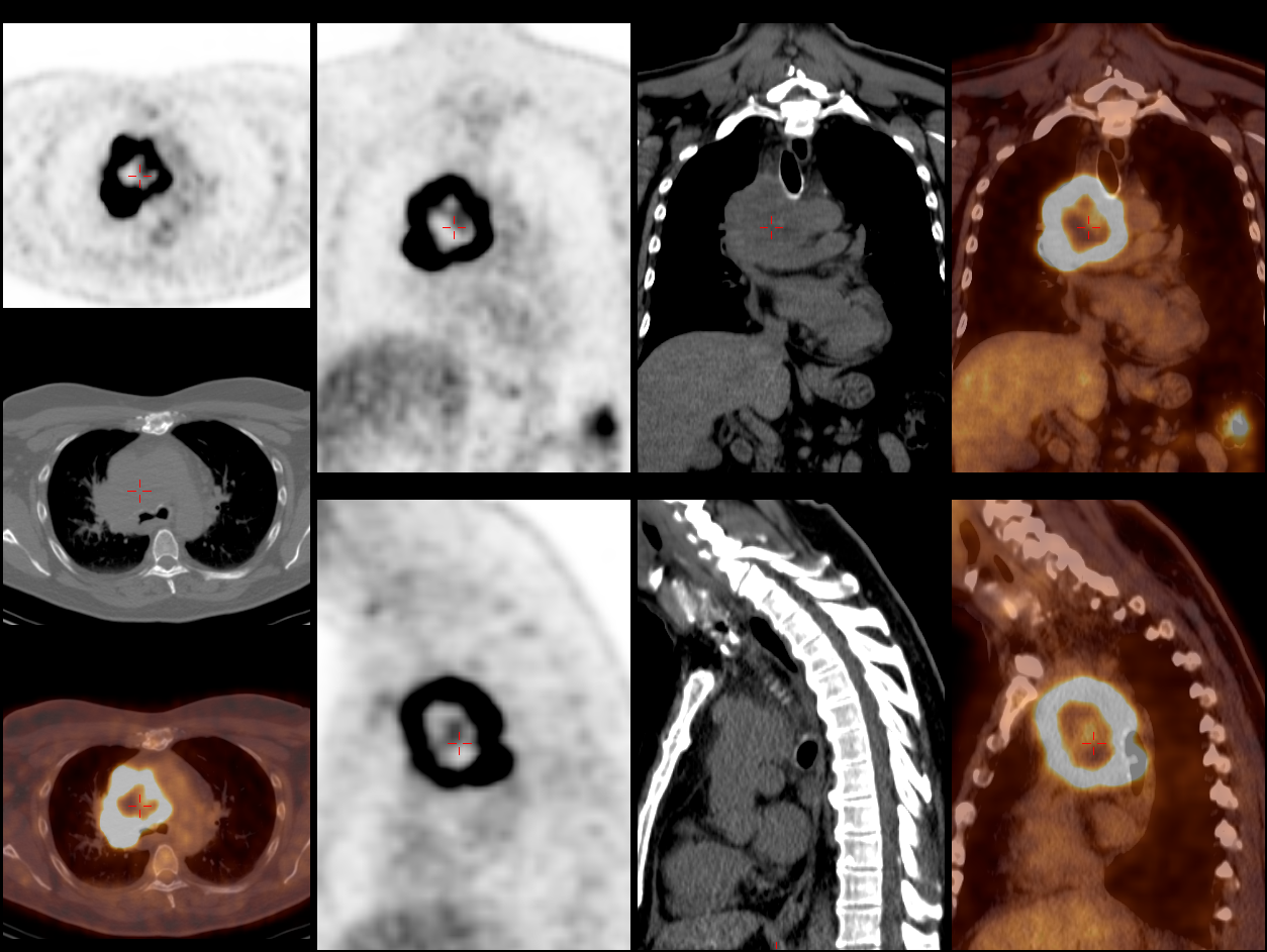
Fig.
6:
PET-CT
scan of the lungs with a tumour necrosis.
For similar reasons to SPECT, PET data can be registered and combined to CT data (see Fig. 6).
Artificial Evolution and Genetic Algorithm
Artificial evolution is a computer science speciality that has emerged in the 70s. It was initially used as a stochastic optimisation technique. It is based on Darwin's observations and on the modern evolution theory, including natural selection, transmission of hereditary properties, and random mutations.
Fig. 7 shows a basic model of a genetic algorithm, one of the main technique in artificial evolution. From an initial population (population of parents), couples of parents are selected to reproduce. Cross-over and mutation are possible to give children. Until the evolution is stopped, children are selected to become parents and so on. This basic model can be modified to match the requirements of the problem to solve.
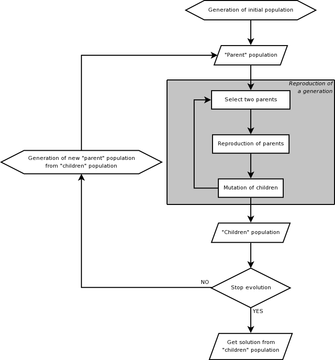
Fig. 7: Basic genetic algorithm.
There are many examples of the use of artificial evolution in image processing and computer vision. It includes image filtering, segmentation, pattern recognition, etc.
Fly Algorithm
The fly algorithm makes use of artificial evolution. Its originality is to consider a fly as a partial solution of the problem to solve, i.e. in our case the reconstructed tomographic volume. A fly corresponds to a point in the 3D space. Depending on the imaging modality, a fly is a photon or positron emitter. Each fly is assessed to evaluate its contribution in the projected images with respect to the original medical data.
Current project:
Fly4PET: Fly Algorithm in PET Reconstruction for Radiotherapy Treatment Planning
References
[3] E. M. Bocage. Technique and mechanism of a moving X-ray film. French patent No. 536464, French Patent Office, Paris, France, 1921. Quoted in [4].
[4] G. W. Friedland and B. O. Thurber. The birth of CT. American Journal of Roentgenology, 167:1365-1370, 1996. Available here (accessed 26th January 2009).
[5] G. N. Hounsfield. Computerized transverse axial scanning (tomography): Part 1. description of system. The British journal of radiology, 46:1016-1022, 1973. PMID: 4757352.
[6] A. C. Kak and M. Slaney. Principles of computerized tomographic imaging. Society of Industrial and Applied Mathematics, 2001. ISBN: 089871494X. Electronic copy available online at http://www.slaney.org/pct/ (accessed 26th January 2009).
[7] R. D. Badawi. Nuclear medicine. Physics Education, 36(6):452-459, November 2001. DOI: 10.1088/0031-9120/36/6/302.